Johns Hopkins researchers report that two brain regions—the entorhinal cortex and amygdala—may begin losing tissue before the hippocampus, which doctors commonly examine for Alzheimer’s-related damage. The changes could be detectable as much as a decade before memory symptoms appear, according to university-provided material published by ScienceDaily on November 14. Source
That’s a potentially valuable head start, but it isn’t a ten-year forecast for an individual. The research identifies possible early biomarkers, or measurable biological indicators. It does not establish a validated clinical test that can predict Alzheimer’s years in advance. Source
Looking beyond the hippocampus
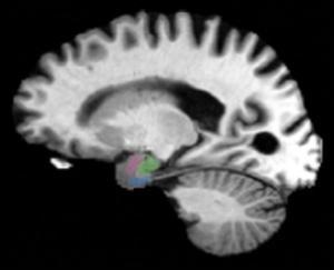
The hippocampus plays a central role in memory and has traditionally received much of the attention when doctors assess Alzheimer’s-related brain damage. The new findings make a case for looking next door, too: the entorhinal cortex and amygdala sit nearby. The former helps retrieve stored memories; the latter contributes to emotional memory. Source
According to the report, the investigation drew on hundreds of magnetic resonance imaging (MRI) and positron emission tomography (PET) scans collected through two research projects: the Alzheimer’s Disease Neuroimaging Initiative and the Biomarkers of Cognitive Decline Among Normal Individuals study. The scans followed people at different disease stages over time, including those without symptoms and those with cognitive impairment. Source
The team reported the same pattern in data from both projects: during early disease stages, the entorhinal cortex and amygdala showed greater relative tissue loss than the hippocampus. Source
“We have found that there are early changes in the brain manifesting the clinical biomarkers of atrophy as much as a decade before there are clinical symptoms,” senior author and biomedical engineer Michael Miller said in the ScienceDaily account. Source
A link to tau, with important limits
Using PET imaging and digital pathology, the researchers also found that areas of gray-matter shrinkage overlapped with regions showing changes associated with tau, a protein that can accumulate abnormally in Alzheimer’s. That supports an association; it does not establish that tau caused the observed tissue loss. Source
The report says the findings were published in NeuroImage: Reports and were being presented at Neuroscience 2026. The account does not specify how many people were studied, how large the tissue-loss differences were or how accurately these changes could predict later symptoms. Source
The researchers next want to investigate molecular changes around abnormal tau accumulation, work they hope could eventually inform new therapies. Source


Comments